Drug Eruptions
- Author: Jonathan E Blume, MD; Chief Editor: Dirk M Elston
Practice Essentials
Drug eruptions can mimic a wide range of dermatoses. The morphologies are myriad and include morbilliform, urticarial, papulosquamous, pustular, and bullous. Medications can also cause pruritus and dysesthesia without an obvious eruption. A drug-induced reaction should be considered in any patient who is taking medications and who suddenly develops a symmetric cutaneous eruption.
Signs and symptoms
The first steps in the history are as follows:
- Review the patient’s complete medication list, including prescription and over-the-counter drugs
- Document any history of previous adverse reactions to drugs or foods
- Consider alternative etiologies (eg, viral exanthems and bacterial infections)
- Note any concurrent infections, metabolic disorders, or immunocompromise
In addition, the following should be noted and detailed:
- Interval between introduction of a drug and onset of the eruption
- Route, dose, duration, and frequency of drug administration
- Use of parenterally administered drugs (more likely to cause anaphylaxis)
- Use of topically applied drugs (more likely to induce delayed-type hypersensitivity)
- Use of multiple courses of therapy and prolonged administration (risk of allergic sensitization)
- Any improvement after drug withdrawal and any reaction with readministration
Physical examination should address clinical features that may indicate a severe, potentially life-threatening drug reaction, including the following:
- Mucous membrane erosions
- Blisters
- Nikolsky sign
- Confluent erythema
- Angioedema and tongue swelling
- Palpable purpura
- Skin necrosis
- Lymphadenopathy
- High fever, dyspnea, or hypotension
It is important to appreciate the morphology and physical features of drug eruptions, as follows:
- Acneiform
- Acral erythema (erythrodysesthesia)
- AGEP
- Dermatomyositislike
- DRESS
- Erythema multiforme (EM), including EM minor, SJS, TEN, and SJS/TEN overlap
- Erythema nodosum
- Erythroderma
- Fixed drug eruptions
- Hypersensitivity syndrome
- Leukocytoclastic vasculitis
- Lichenoid
- Lupus
- Morbilliform or exanthematous
- Pseudoporphyria[1]
- Serum sickness and serum sickness–like
- Sweet syndrome (acute febrile neutrophilic dermatosis)
- Urticaria
- Vesiculobullous
See Clinical Presentation for more detail.
Diagnosis
With mild asymptomatic eruptions, the history and physical examination are often sufficient for diagnosis; with severe or persistent eruptions, further diagnostic testing may be required, as follows:
- Biopsy
- Complete blood count (CBC) with differential
- Serum chemistry studies (especially for electrolyte balance and indices of renal or hepatic function in patients with severe reactions)
- Antibody or immunoserology tests
- Direct cultures to investigate a primary infectious etiology or secondary infection
- Urinalysis, stool guaiac tests, and chest radiography for vasculitis
- Skin prick or patch testing to confirm the causative agent
See Workup for more detail.
Management
Principles of medical care are as follows:
- The ultimate goal is to identify and discontinue the offending medication if possible
- Patients can sometimes continue to be treated through morbilliform eruptions; nevertheless, all patients with severe morbilliform eruptions should be monitored for mucous membrane lesions, blistering, and skin sloughing
- Treatment of a drug eruption depends on the specific type of reaction
- Therapy for exanthematous drug eruptions is supportive, involving the administration of oral antihistamines, topical steroids, and moisturizing lotions
- Severe reactions (eg, SJS, TEN, and hypersensitivity reactions) warrant hospital admission
- TEN is best managed in a burn unit, and intravenous immunoglobulin (IVIG) may improve outcomes[2, 3, 4]
- Hypersensitivity syndrome may have to be treated with liver transplantation if the offending drug is not stopped in time; treatment with systemic corticosteroids has been advocated in the acute phase; in the chronic phase, patients may require treatment for hypothyroidism or diabetes mellitus
For most drug eruptions, full recovery without any complications is expected; however, the following should be noted:
- Patients with exanthematous eruptions should expect mild desquamation as the rash resolves
- Patients with hypersensitivity syndrome are at risk of becoming hypothyroid, usually within the first 4-12 weeks after the reaction; there is also a risk of diabetes
- The prognosis for patients with TEN is guarded; scarring, blindness, and death are possible
Image library
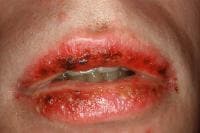 Stevens-Johnson syndrome.
Background
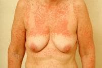
Drug eruptions can mimic a wide range of dermatoses. The morphologies are myriad and include morbilliform (most common, see image below), urticarial, papulosquamous, pustular, and bullous. Medications can also cause pruritus and dysesthesia without an obvious eruption. Both calcium channel blockers and interferon are strongly associated with eczematous eruptions.
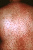 Morbilliform drug eruption.
A drug-induced reaction should be considered in any patient who is taking medications and who suddenly develops a symmetric cutaneous eruption. Medications that are known for causing cutaneous reactions include antimicrobial agents,[5] nonsteroidal anti-inflammatory drugs (NSAIDs), cytokines, chemotherapeutic agents, anticonvulsants, and psychotropic agents.
Prompt identification and withdrawal of the offending agent may help limit the toxic effects associated with the drug. The decision to discontinue a potentially vital drug often presents a dilemma.
Pathophysiology
Drug eruptions may be divided into immunologically and nonimmunologically mediated reactions.
Immunologically mediated reactions
Coombs and Gell proposed 4 types of immunologically mediated reactions, as follows:
- Type I is immunoglobulin E (IgE)–dependent reactions, which result in urticaria, angioedema, and anaphylaxis (see the image below).
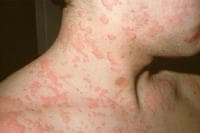 Urticaria.
 - Type II is cytotoxic reactions, which result in hemolysis and purpura (see the image below).
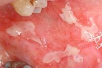 Oral ulcerations in a patient receiving cytotoxic therapy.
 - Type III is immune complex reactions, which result in vasculitis, serum sickness, and urticaria.
- Type IV is delayed-type reactions with cell-mediated hypersensitivity, which result in contact dermatitis, exanthematous reactions, and photoallergic reactions.
Th17 T cells are implicated in many drug eruptions, and sulfamethoxazole induces a T-cell switch mechanism based on the TCRVβ20-1 domain altering peptide-HLA recognition. In severe drug reactions, micro RNA-18a-5p down-regulates the expression of the antiapoptotic B-cell lymphoma/leukemia-2–like protein 10 (BCL2L10), promoting apoptosis.
Insulin and other proteins are associated with type I reactions. Penicillin, cephalosporins, sulfonamides, and rifampin are known to cause type II reactions. Quinine, salicylates, chlorpromazine, and sulfonamides can cause type III reactions. Type IV reactions, the most common mechanism of drug eruptions, are often encountered in cases of contact hypersensitivity to topical medications, such as neomycin. Sulfonamides are most frequently associated with toxic epidermal necrolysis (TEN).
Although most drug eruptions are type IV hypersensitivity reactions, only a minority are IgE-dependent. That is, antibodies can be demonstrated in less than 5% of cutaneous drug reactions. Type IV cell-mediated reactions are not dose dependent, they usually begin 7-20 days after the medication is started, they may involve blood or tissue eosinophilia, and they may recur if drugs chemically related to the causative agent are administered.
Nonimmunologically mediated reactions
Nonimmunologically mediated reactions may be classified according to the following features: accumulation, adverse effects, direct release of mast cell mediators, idiosyncratic reactions, intolerance, Jarisch-Herxheimer phenomenon, overdosage, or phototoxic dermatitis. (Symptoms of Jarisch-Herxheimer reactions disappear with continued therapy. Drug therapy should be continued until the infection is fully eradicated.)
An example of accumulation is argyria (blue-gray discoloration of skin and nails) observed with use of silver nitrate nasal sprays.
Adverse effects are normal but unwanted effects of a drug. For example, antimetabolite chemotherapeutic agents, such as cyclophosphamide, are associated with hair loss.
The direct release of mast cell mediators is a dose-dependent phenomenon that does not involve antibodies. For example, aspirin and other NSAIDs cause a shift in leukotriene production, which triggers the release of histamine and other mast-cell mediators. Radiographic contrast material, alcohol, cytokines, opiates, cimetidine, quinine, hydralazine, atropine, vancomycin, and tubocurarine also may cause release of mast-cell mediators.
Idiosyncratic reactions are unpredictable and not explained by the pharmacologic properties of the drug. An example is the individual with infectious mononucleosis who develops a rash when given ampicillin.
Imbalance of endogenous flora may occur when antimicrobial agents preferentially suppress the growth of one species of microbe, allowing other species to grow vigorously. For example, candidiasis frequently occurs with antibiotic therapy.
Intolerance may occur in patients with altered metabolism. For example, individuals who are slow acetylators of the enzyme N -acetyltransferase are more likely than others to develop drug-induced lupus in response to procainamide.
Jarisch-Herxheimer phenomenon is a reaction due to bacterial endotoxins and microbial antigens that are liberated by the destruction of microorganisms. The reaction is characterized by fever, tender lymphadenopathy, arthralgias, transient macular or urticarial eruptions, and exacerbation of preexisting cutaneous lesions. The reaction is not an indication to stop treatment because symptoms resolve with continued therapy. This reaction can be seen with penicillin therapy for syphilis, griseofulvin or ketoconazole therapy for dermatophyte infections, and diethylcarbamazine therapy for oncocerciasis.
Overdosage is an exaggerated response to an increased amount of a medication. For example, increased doses of anticoagulants may result in purpura.
Phototoxic dermatitis is an exaggerated sunburn response caused by the formation of toxic photoproducts, such as free radicals or reactive oxygen species (see the image below).
 Phototoxic reaction after use of a tanning booth. Note sharp cutoff where clothing blocked exposure.
Frequency
United States
Drug eruptions occur in approximately 2-5% of inpatients and in greater than 1% of outpatients.
International
Drug eruptions occur in approximately 2-3% of inpatients.
Mortality/Morbidity
Most drug eruptions are mild, self-limited, and usually resolve after the offending agent has been discontinued. Severe and potentially life-threatening eruptions occur in approximately 1 in 1000 hospital patients. Mortality rates for erythema multiforme (EM) major are significantly higher. Stevens-Johnson syndrome (SJS) has a mortality rate of less than 5%, whereas the rate for TEN approaches 20-30%; most patients die from sepsis.
Sex
Adverse cutaneous reactions to drugs are more prevalent in women than in men.
Age
Elderly patients have an increased prevalence of adverse drug reactions